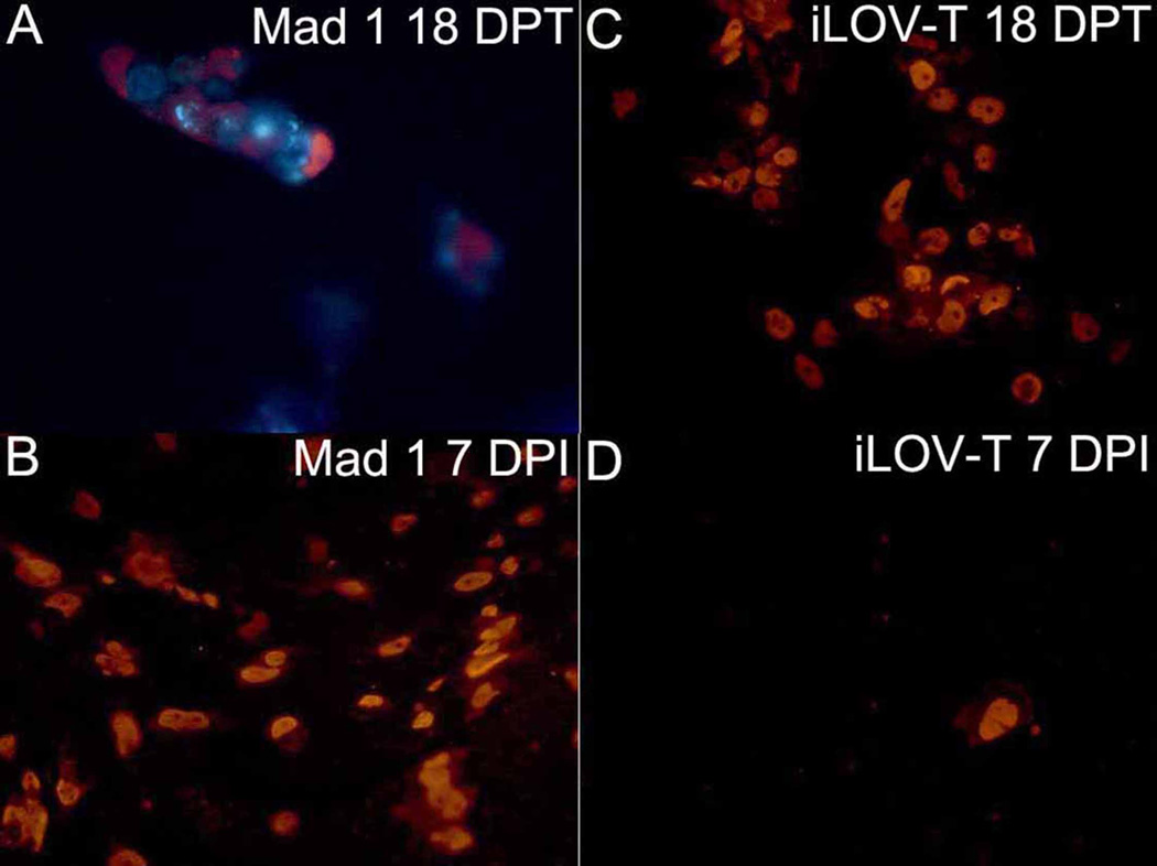
Fig. 4.
Intracellular staining (ICS) of JC virus VP1 protein in 293FT cells transfected or infected with JCV Mad1 (A,B) or JCV iLOV-T (C,D). JCV VP1 ICS is performed as described previously, using fluorescent reporter Alexa Fluor 568. 293FT cells 18 days post transfection (upper panels) or 7 days post infection (lower panels) are shown. Cells that are red express JCV VP1 protein

