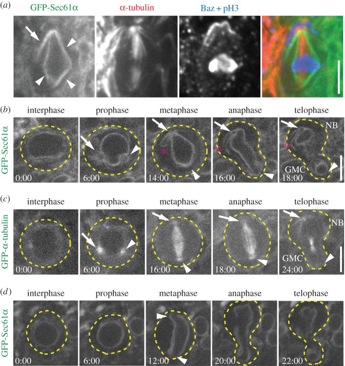
Figure 1.
The ER is asymmetrically partitioned in dividing NBs. (a) A fixed metaphase NB expressing GFP-Sec61α (green) was immunostained for α-tubulin (red), Baz (blue) and phosphorylated histone 3 (pH3, blue). Indicated are the ER envelope (arrowheads) and the apical ER extension (arrow). (b) A single GFP-Sec61α expressing NB was imaged live throughout the course of a single mitotic division. Depicted are the approximate outline of the cell (dotted lines), apical centrosome (arrow), the basal centrosome (arrowhead) and the apical ER extension (asterisk). (c) A single GFP-α-tubulin expressing NB was imaged live throughout the course of a single mitotic division. The apical centrosome (arrow) and the basal centrosome (arrowhead) are shown. (d) An aslmecD/aslmecD, GFP-Sec61α expressing NB was imaged live throughout the course of a single mitotic division. Indicated are the approximate outline of the cell (dotted lines) and the spindle poles (arrow heads). Note the lack of centrosomal and spindle pole accumulations of ER. Times, min:s; scale bar, 5 µm.

