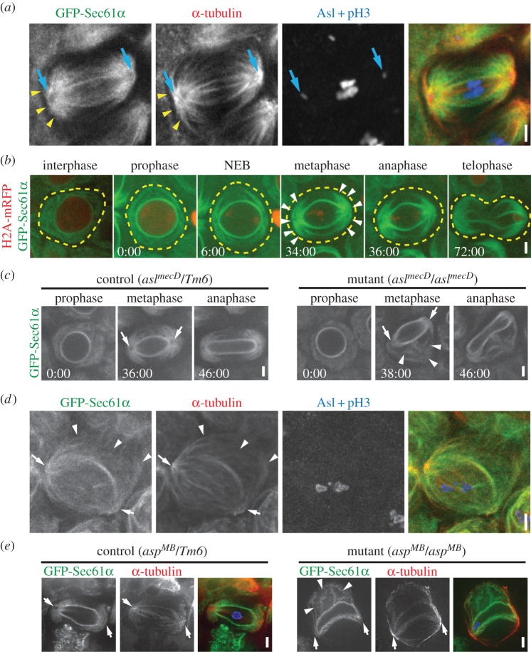
Figure 2.
The ER associates with astral MTs in meiotic spermatocytes. (a) A fixed GFP-Sec61α (green) spermatocyte at metaphase of the first meiotic division was immunostained for α-tubulin (red), asterless (Asl, blue) to localize centrosomes (blue arrows) and phosphorylated histone 3 (pH3, blue). ER structures are closely aligned with astral MTs (yellow arrowheads). (b) A spermatocyte expressing GFP-Sec61α (green) and H2A-mRFP (red) was imaged live throughout the course of the first meiotic division. NEB was determined based on the sudden loss of background H2A-mRFP fluorescence throughout the nucleus. The approximate outline of the cell (yellow dotted lines) and the astral ER domains at metaphase (arrow heads) are indicated. (c) aslmecD/TM6 control (left panel) and aslmecD/aslmecD (right panel) GFP-Sec61α expressing spermatocytes were imaged throughout meiosis I. Spindle poles (arrows) recruit ER in control cells, but fail to do so in aslmecD/aslmecD spermatocyte; abnormal ER structures (arrowheads) are prominent in the aslMecD/aslMecD spermatocyte. (d) A fixed GFP-Sec61α (green) metaphase I aslMecD/aslMecD spermatocyte was immunostained for α-tubulin (red), asl (blue) and pH3 (blue). Indicated are spindle poles (arrows) and abnormal ER structures associated with MTs (arrowheads). (e) Fixed GFP-Sec61α (green) metaphase I aspMB/TM6 control (left panel) and aspMB/aspMB (right panel) spermatocytes were fixed and immunostained for α-tubulin (red). DAPI staining of DNA is shown in blue in the merged image. Abnormal ER structures are prominent in aspMB/aspMBspermatocyte (arrows). Times, min:s; scale bar, 5 µm.

