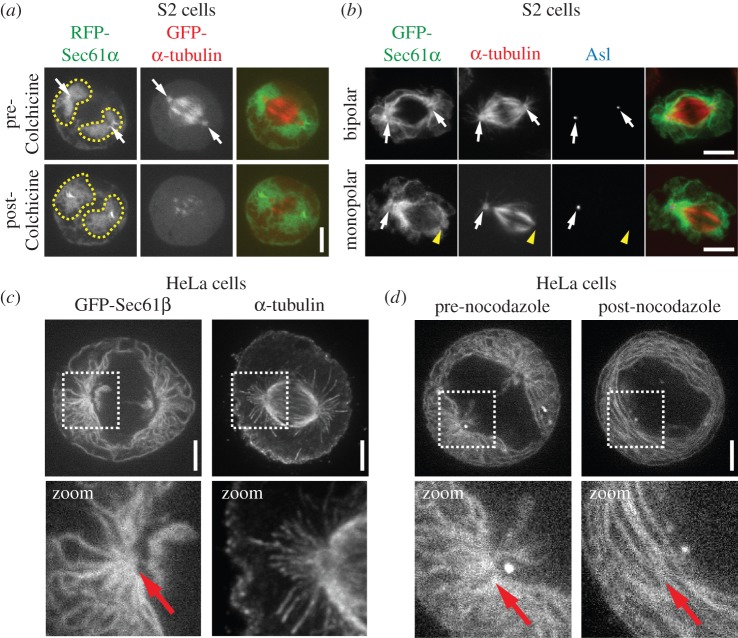
Figure 4.
The ER is organized by spindle poles and astral MTs in human and Drosophila tissue culture cells. (a) A metaphase Drosophila S2 cell expressing RFP-Sec61α (green) and GFP-α-tubulin (red) was imaged live pre- and post-treatment with 100 µM colchicine. The post-colchicine images were taken 20 min following application of the drug. Spindle poles (arrows) and the ER domains surrounding the spindle poles (dotted lines) are indicated. (b) GFP-Sec61α (green) expressing S2 cells were fixed and stained for α-tubulin (red) and Asl (blue). The top panel shows a normal metaphase cell with two centrosomes, marked by Asl (arrows) at both spindle poles. The bottom panel shows a metaphase cell with only a single centrosome located at one of the spindle poles (arrow); the other spindle pole is acentrosomal (yellow arrow head). Note that the acentrosomal spindle pole completely lacks astral MTs and is associated with much less ER than the centrosome-containing pole. (c) Shown on the left is a live metaphase HeLa cell expressing GFP-Sec61β, and shown on the right is a fixed metaphase HeLa cell stained for α-tubulin to show the organization of a typical spindle in these cells. Note the radial arrays of ER that are organized around the spindle poles and extend towards the cell cortex, and the resemblance of these arrays to the astral MTs seen in the α-tubulin image (highlighted in zoomed images below). (d) A metaphase GFP-Sec61β expressing HeLa cell was imaged live pre- and post-treatment with 10 µM nocodazole. The post-treatment image was taken 20 min following application of the drug. A clear loss of the ER radial array can be seen following MT depolymerization (highlighted in zoomed images below). Scale bars, 5 µm.

