Figure 2.
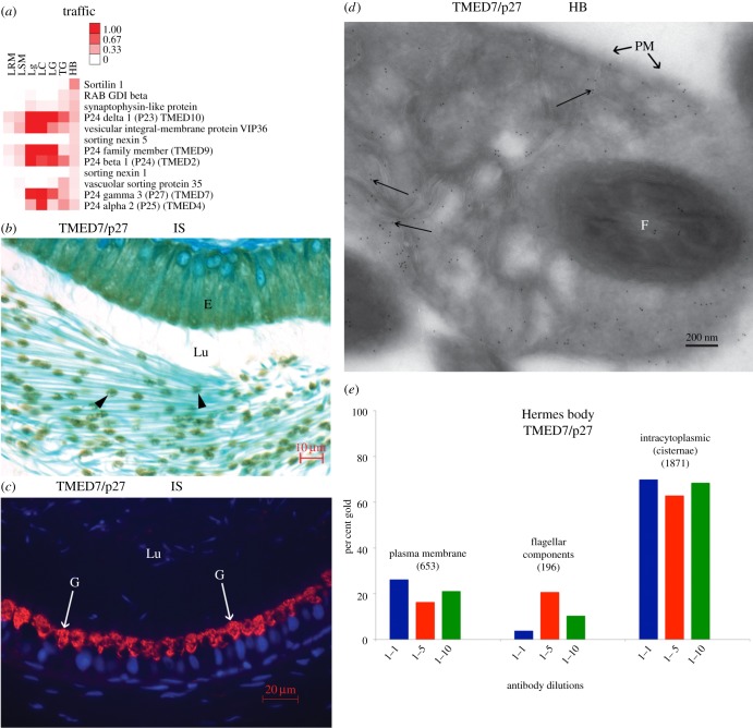
Hermes bodies localization of TMED7/p27 by LM and EM in situ. (a) Heat map of the 12 most abundant proteins of the Traffic category. (b) IHC shows the localization of TMED7/p27 in epididymal Hermes bodies. (c) IF reveals Golgi reactivity of epididymal epithelial cells for TMED7/p27. However, germ cell immunoreactivity is not seen due to the low sensitivity of the IF protocol. (d) EM immunolocalization of TMED7/p27. Gold particles (arrows) over flattened cisternae, plasma membrane and flagella of sperm. (e) Gold particle quantification in cryosections of Hermes body of sperm. The total number of gold particles scored over the plasma membrane, intracytoplasmic (cisternae) or flagella are indicated above each histogram representing different dilutions of anti-TMED7/p27. E, epithelium; F, flagellum; G, Golgi reactivity; HB, Hermes body; IS, initial segment; Lu, lumen; PM, plasma membrane.

