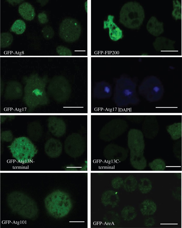
Figure 2.
Cellular localization of Atg1 complex proteins by confocal microscopy. For comparison, the autophagic marker GFP-Atg8 is included, which shows the typical autophagosome punctated pattern. Atg101, Atg13-C-terminal, Atg13-N-terminal, FIP200 and Atg17-GFP tagged proteins were expressed and visualized in vivo by confocal microscopy. GFP-Atg17 expressing cells were also stained with DAPI for co-localization with nuclei. Scale bars, 10 µm.

