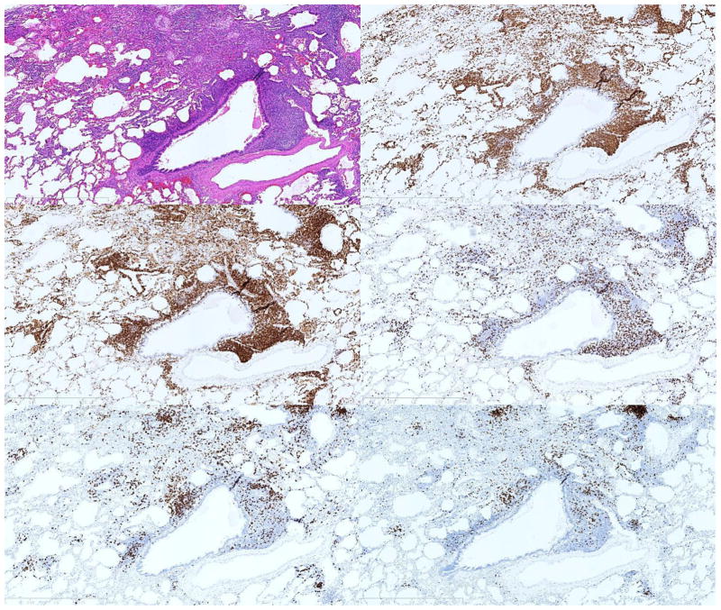
Figure F–K.
T lymphocyte predominance in GLILD. FB pattern infiltrate in GLILD (F) with predominantly CD3 positive (G), CD4 positive T cells (H), with smaller numbers of CD8 positive T cells (I) and CD20 positive (J), PAX-5 positive B cells (K). This pattern of T lymphocyte predominance was commonly observed.

