Figure 1.
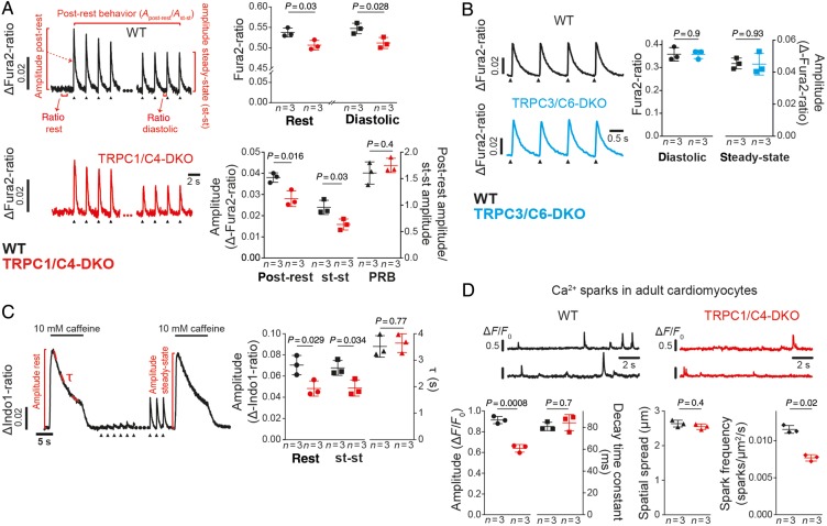
Ca2+ signalling in cardiomyocytes from TRPC1/C4-DKO is reduced. (A) Representative electrically evoked Ca2+ transients (0.5 Hz, arrow heads) in adult ventricular myocytes. Here resting Ca2+ (rest) and diastolic Ca2+ levels during steady-state (st-st) pacing, and amplitudes of Ca2+ transients post-rest and during steady-state pacing are depicted. The post-rest behaviour, PRB, (post-rest amplitude/st-st amplitude) is shown (44–55 cells/group). (B) Analysis of electrically evoked Ca2+ transients (1 Hz) in TRPC3/C6-DKO myocytes (119–191 cells/group). (C) Analysis of caffeine-induced Ca2+ transients in resting cardiomyocytes or immediately after a 3-min pacing period (st-st conditions). Typical transients are depicted highlighting the amplitudes (65–75 cells/group) and decay time constant (τ) (52–68 cells/group). (D) Recordings of two different spark sites and the corresponding analysis of decay time constant, spatial spread and frequency (24–32 cells/group).

