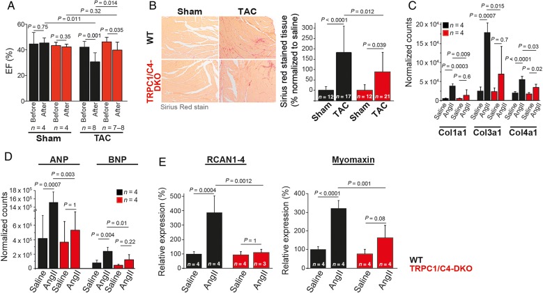
Figure 5.
Reduced pathological cardiac hypertrophy in TRPC1/C4-DKO mice. (A) Ejection fraction (EF) measured by echocardiography before and 5 weeks after TAC. (B) Reduced ventricular fibrosis in TRPC1/C4-DKO mice after TAC. Expression analysis of collagen genes (C), ANP and BNP (D), and the reporters of Ca2+-dependent hypertrophy signalling through NFAT and MEF2, RCAN and Myomaxin (E) after AngII infusion.

