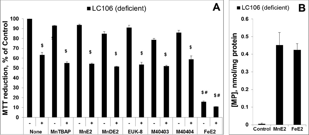
Figure 8. Evaluation of metal complexes in an E. coli model (catalase/peroxidase-deficient LC106 strain) of H2O2-induced damage – drugs were pre-incubated with E. coli prior to H2O2 addition.
Except Mn salen, EUK-8, all other Mn complexes studied are the same as those in Figure 7. Two new abbreviations are introduced, MnE2 being MnTE-2-PyP5+ and MnDE2 being MnTDE-2-ImP5+. After 1 hour pre-incubation of E. coli with 20 µM of the drugs, the catalase/peroxidase mutant (LC106) cells were washed with PBS and exposed to 0.5 mM H2O2. After 15 min of incubation, H2O2 was decomposed by adding 1,000 units/ml of catalase (A). The viability was measured via MTT test and expressed as a percentage of the MTT reduction by non-treated cells. None of the compounds were toxic, but were also not able to suppress H2O2 toxicity. Fe porphyrin, FeTE-2-PyP5+, was toxic under given conditions. To verify the presence of pentacationic porphyrins in cells, the accumulation of MnTE-2-PyP5+ vs FeTE-2-PyP5+ during 1 hour of incubation was determined and appeared similar (plot B). Student t-test was used to determine the statistical significance. Mean ± S.E is presented. $statistical significance (p<0.05) compared to untreated cells; # statistical significance (p<0.05) compared to cells treated with H2O2 only.

