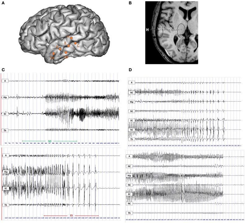
Figure 1.
Intracerebral recordings using multiple contacts electrodes placed according to Talairach’s stereotactic method. (A) Schematic diagram of SEEG electrodes placement on a lateral view of a three-dimensional (3D) reconstruction of the neocortical surface in a patient with MTLE. Electrode A explored the amygdala (internal leads) and the anterior part of the MTG (lateral leads); electrode B explored the anterior hippocampus (internal leads) and the middle part of MTG (lateral leads); electrode TB explored the entorhinal cortex (internal leads) and the anterior part of ITG (external leads); electrode C explored the posterior hippocampus (internal leads) and the posterior part of MTG (lateral leads); electrode H explored the thalamus (internal leads) and the posterior part of STG (lateral leads). (B) MRI axial view of standard orthogonal electrode H exploring thalamus by its internal leads and STG by its external leads. (C) Ictal SEEG recordings with selected traces from mesio-temporal structures, i.e., amygdala (A), hippocampus (Hip), entorhinal cortex (EC), and from thalamus (Th). Seizure onset period (SO) included 5 s before and 10 s after the onset of high-frequency activity in mesial temporal structures (top panel); the end of seizure period (ES) was defined as the last 15 s of the seizure discharge (bottom panel). (D) SEEG recordings of two seizures in the ES period displaying the thalamic rhythmic pattern with a spike-wave activity (on the top) or an arched morphology (on the bottom) coincidental with cortical spike-and-wave discharges. A, amygdala; Hip, hippocampus; EC, entorhinal cortex; NC, neocortex; Th, thalamus.

