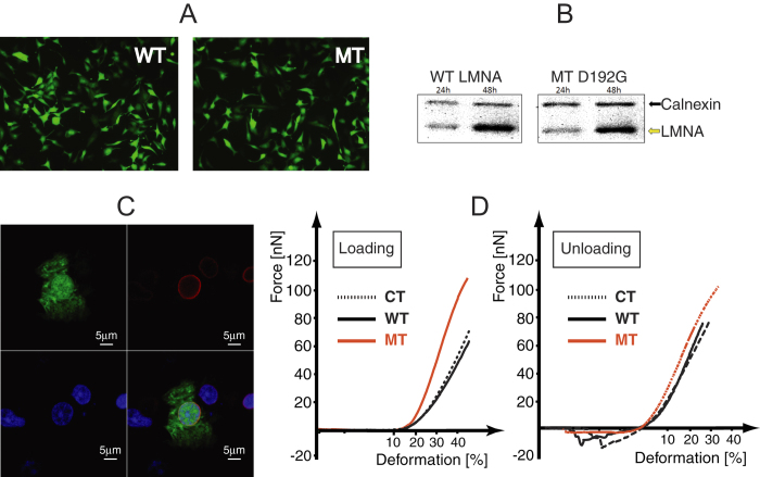
Figure 1.
(A) EGFP and human LMNA expression detected by fluorescence light microscopy 24 hours post-infection with adenoviral WT and MT NRVM’s constructs. (B) Expression of the transduced human LMNA protein detected by human specific anti-LMNA antibody by western blot: human LMNA and rat Calnexin at 24 and 48 hours post-infection. (C) Indirect immunofluorescence showing co-localization between GFP (green, upper-left panel) and human LMNA (red, right-upper panel) signals in D192G LMNA NRVMs, transduced by adenoviral bicistronic GFP-LMNA construct. The nuclei are stained in blue (TOTO3, lower-left column). The fluorescence is represented in three channels. Bars: 5 μ. As seen in the merged panel (lower-right), human LMNA is perfectly co-localized in the area of the nuclear wall. (D) AFM loading-unloading curves for CT, WT LMNA and MT LMNA D192G.

