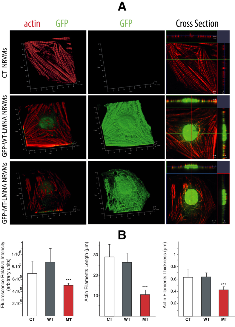
Figure 3. D192G LMNA seems to induce actin network alteration in NRVMs:
(A) 3D reconstruction of the appearance of actin cytoskeleton (red) in NRVMs in control conditions and after infection with WT and MT-GFP LMNA. NRVMs infected with MT-GFP LMNA displayed a lower actin fibers density into the cytoskeleton and the presence of blebs on nuclear membrane clearly visible in a 3D reconstruction of a Z-stack acquired in a range of 9.81 μm. (B) Quantification of red fluorescence relative intensity (n = 3 optical fields for each experimental condition), and appearance of labeled actin filaments length (CT n = 38 fibers; WT n = 40 fibers; MT n = 67 fibers) and thickness (CT n = 67 fibers; WT n=86 fibers; MT n = 82 fibers). ***Student t-Test significant difference in comparison to controls (CT) at p < 0.001. Data show a significant decrease of fluorescence relative intensity, actin filaments length and actin filaments thickness in MT NRVM compared to CT and WT cells.

