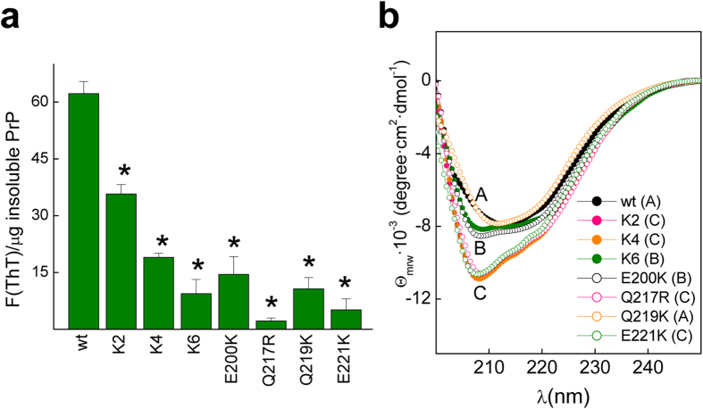
Figure 5. Properties of the PrP wt and mutant fibrils.
(a) Specific ThT binding of the PrP wt and mutant fibrils. Typically 50 μM of fibrils in 10 mM ammonium acetate pH 5 were incubated for 10 min with ThT (15 μM) before fluorescence determination. ThT fluorescence intensities were corrected for the background (absence of fibrils) and divided by the protein amount in the pellet of a 12000 rpm 20 min centrifugation. The depicted data represent the average of two independent experiments performed in duplicate (*p < 0.01). (b) Far-UV CD spectra of the PrP wt and mutant assemblies in 10 mM ammonium acetate pH 5. The fibrils were formed in 50 mM MES pH 6.5 containing 2 M GdnCl at 37 °C under continuous rotation at 24 rpm, and then dialyzed against 10 mM ammonium acetate pH 5.

