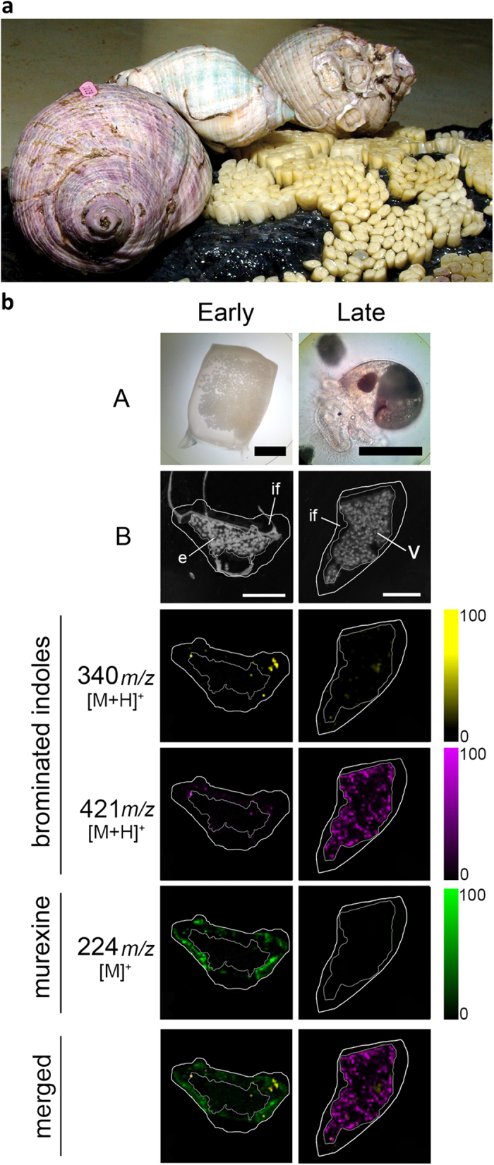
Figure 5. D. orbita during egg deposition and DIOS-MSI maps of egg capsules across the developmental period, in positive ion mode at 100 μm spatial resolution.

(a) Reproductive adults during the encapsulation of larvae and early stage capsules adhered to substrate (photo by Rudd, D.). (b) early stage capsule sampled immediately post deposition (left panels A = whole capsule) and late stage capsule after 35 days post deposition (right panels A = encapsulated veliger larvae). DIOS-MSI of the secondary metabolites are compared to (B) scanned cross sections of the egg capsules stamped onto pSi prior to removal. Labels on the imaged regions include; embryo mass (e), intracapsular fluid, (if) and veligar (v) stage larval mass. Ion maps m/z 340 corresponds to tyrindoxyl hydrogen sulfate [M+H]+, m/z 421 to Tyrian purple [M+H]+, and m/z 224 to murexine [M]+. Scale bar set to 1 mm.
