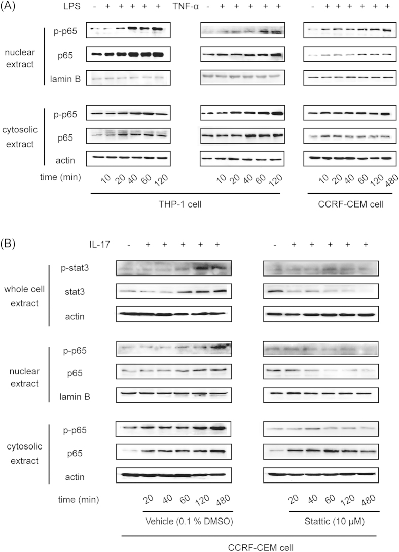
Figure 7. IL-17 increased P-gp expression through the STAT3-dependent Nf-κb pathway.
(A) THP-1 cells were treated with 50 ng/ml TNF-α or 2 μg/ml LPS for different periods of time. CCRF-CEM cells were treated with 50 ng/ml TNF-α for different periods of time. Nuclear and cytosolic p65 and phosphorylated p65 were detected by Western blot. (B) CCRF-CEM cells were treated with 200 ng/ml IL-17 in the absence or presence of the STAT3 inhibitor Stattic for different periods of time. Total STAT3 and phosphorylated STAT3 and nuclear and cytosolic p65 and phosphorylated p65 were detected by Western blot.

