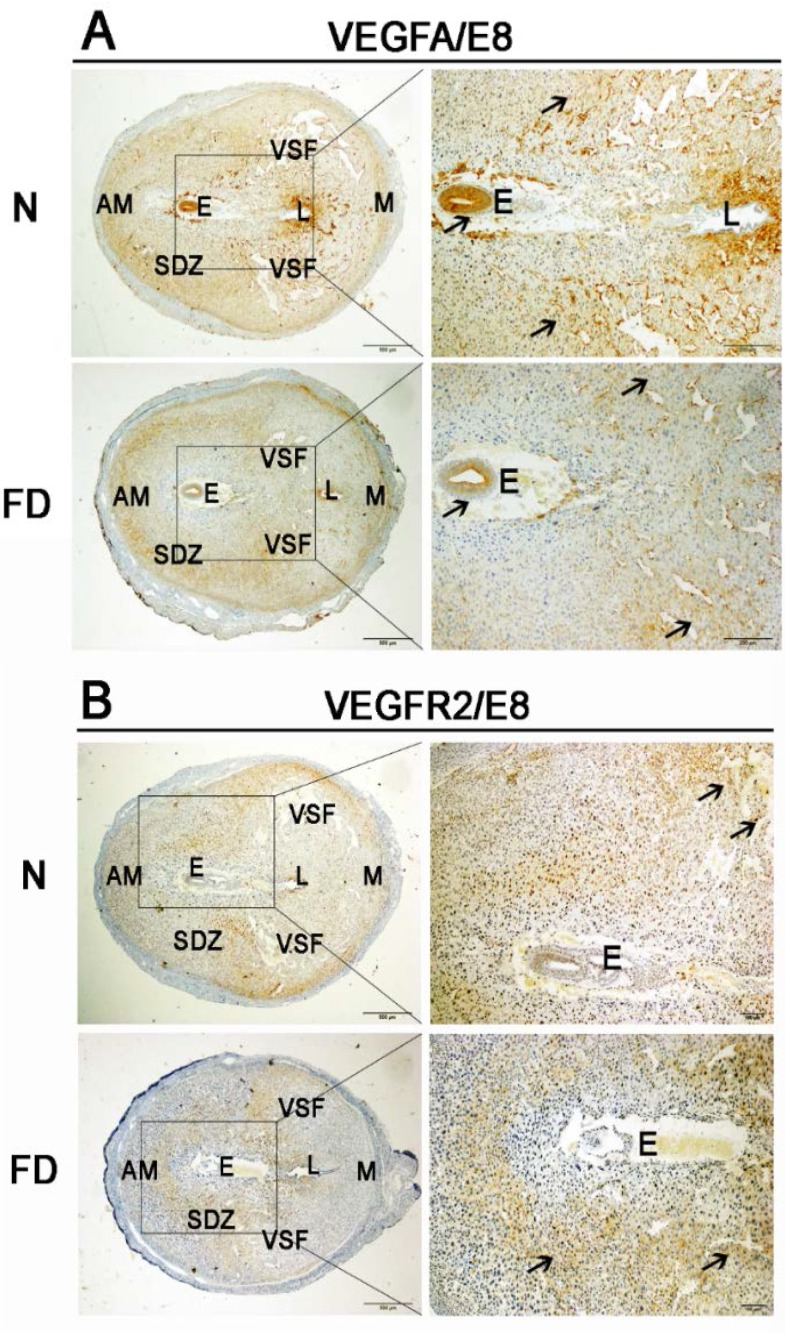
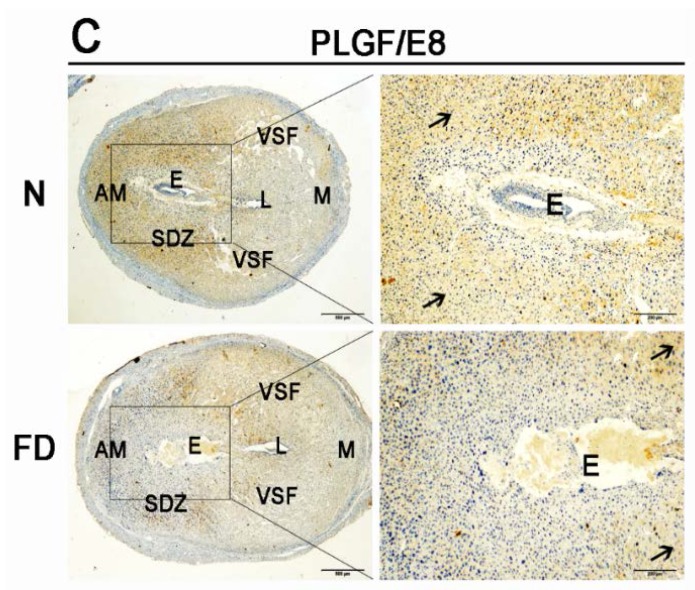
Figure 3.
Immunohistochemistry staining with vascular endothelial growth factor A (VEGFA), vascular endothelial growth factor receptor 2 (VEGFR2), and placental growth factor (PLGF). Decreased expression of VEGFA, VEGFR2, and PLGF was detected in the folate-deficient group. The pictures in the right column are the higher magnification images of the black boxes in the left column. Additionally, the black boxes were the uterus’s central region around the embryo. Arrow indicates each factor’s different expression and distribution between two groups. (A) VEGFA was localized to a wide area of the secondary decidual zone (PDZ) and the embryo in both normal and folate-deficient mice, but there was decreased expression in the folate-deficient group. (B) VEGFR2 was localized mainly to the VSF close to the AM region in normal mice, whereas VEGFR2 was localized mainly to part of the PDZ in folate-deficient mice. (C) PLGF had a similar localization pattern as VEGFA in normal mice but was not expressed in the embryo. However, PLGF was expressed mainly in the central region of the uterus instead of in the PDZ in folate-deficient mice. N: normal group; FD: folate-deficient group; E: embryo; L: luminal epithelium; SDZ: secondary decidual zone; M: mesometrial; AM: anti-mesometrial; VSF: vascular sinus folding. Scale bar: 500 μm (left), 200 μm (right).


