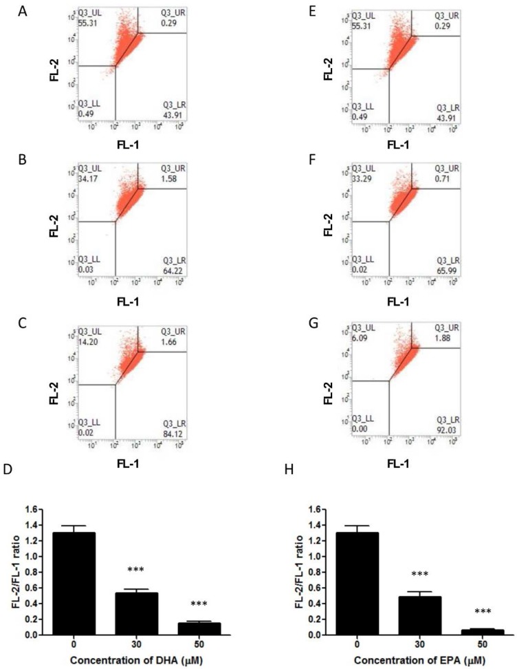
Figure 5.
DHA and EPA reduce mitochondrial membrane potential in LA-N-1 cells. (A–D) LA-N-1 cells were incubated with ethanol control (A), 30 or 50 µM DHA (B, C) for 48 hours. (E–H) LA-N-1 cells were incubated with ethanol control (E), 30 or 50 µM EPA (F, G) for 48 hours. After incubation, the cells were stained by JC-1 dye and the mitochondrial potentials were detected by flow cytometry. The results are quantified and expressed as mean values ± SD (D, H). *** p < 0.001.

