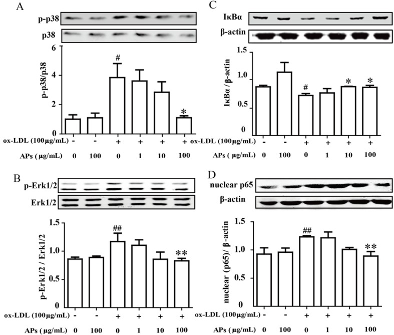
Figure 7.
APs inhibit the MAPK and NF-κB activation induced by ox-LDL in RAECs. RAECs were cultured in media with various concentrations of APs for 1 h, followed stimulation with ox-LDL for 30 min to assess the MAP kinase pathway or for 60 min to assess the NF-κB pathway. A and B: Activation of p38 or Erk1/2 was determined by immunoblotting using phosphor-specific (p-) antibodies. Representative blots from three independent experiments are shown. The quantitation of the ratio between phosphorylated protein/total proteins in the cell lysates is shown in the bar graphs; C and D: The nuclear and cytosolic fractions of NF-κB were extracted and then assessed by Western blot. The results are shown as the means ± S.E.M. # p < 0.05, ## p < 0.01 vs. control group. * p < 0.05, ** p < 0.01 vs. ox-LDL alone.

