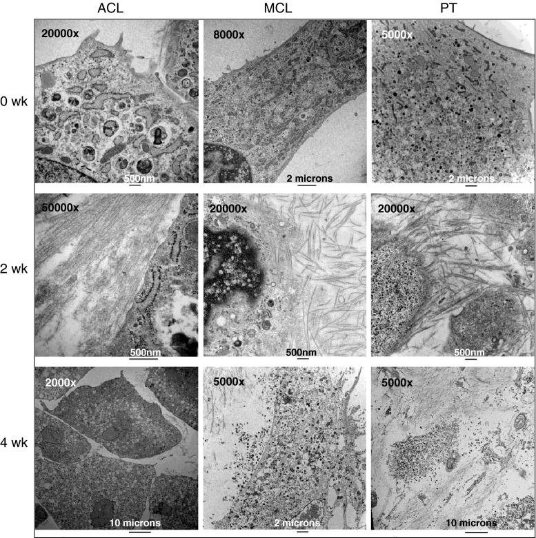
Fig. 4.
Transmission electron microscopy (TEM) photomicrographs showed collagen fibrils secreted from rabbit anterior cruciate ligament (ACL), medial collateral ligament (MCL), and patellar tendon (PT) cells at 0, 2, and 4 weeks after reaching confluence in culture. The TEM photomicrographs showed a random orientation of the deposited fibrous matrix. The cytoplasm of the cells contained large quantities of rough-surfaced endoplasmic reticulum when the cells reached confluence in culture. Two weeks after reaching confluence, all of the cells secreted large amounts of collagen. However, the cytoplasm of ACL cells contained many lacunae. After 4 weeks in culture, the plasma membrane of the MCL and PT cells had ruptured releasing the contents of the cytoplasm from the cells because of deterioration in the conditions of the culture environment

