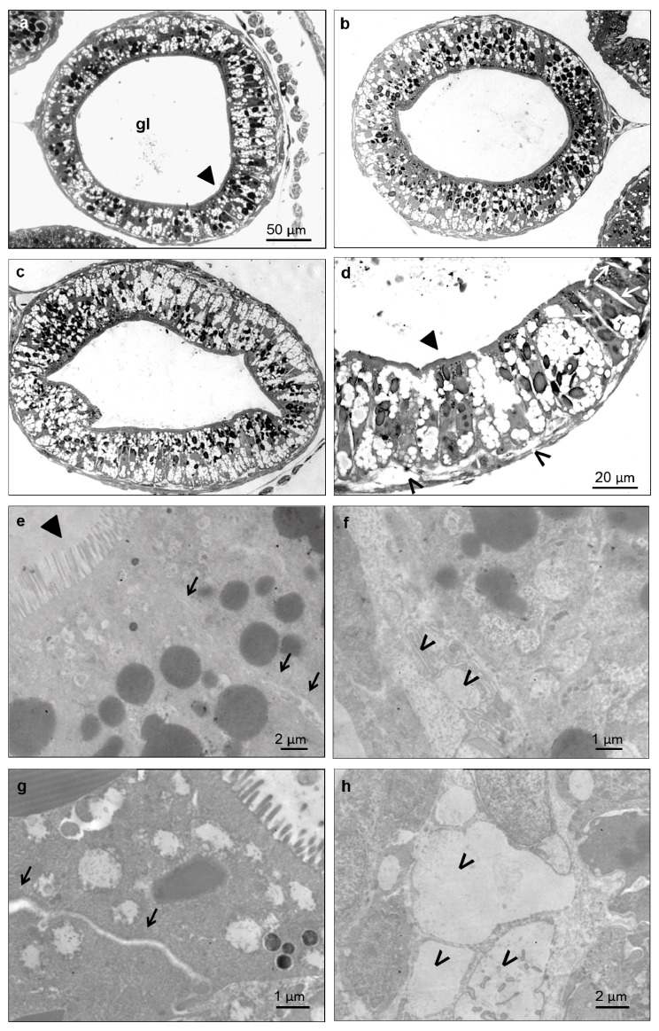
Figure 7.
Light (a–d) and electron microscopy (e–h) imaging of the X. laevis small intestine. Transversal sections at the level of an intestinal loop of a control (a), bZnO (b) and sZnO (c,d) exposed embryos. Magnification of the sZnO intestinal loop (d) shows the swelling of paracellular spaces between cells (empty arrow) and detachment in some regions of epithelial cells from basal lamina (*). These damages are more evident in the detail of the junctional complex between two enterocytes (g) (black arrow) and of the basal portion (h) (*) of sZnO-exposed embryos in comparison to the control (e) (black arrow) and (f) (*). ► = brush border; gl = gut lumen.

