Figure 1.

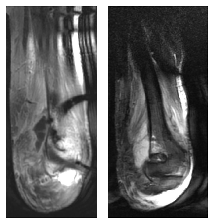
MRI (coronal T1 and STIR) showing a collection of 51 × 23 mm with bone edema in the humerus supratrochlear region with apparent cortical integrity.

MRI (coronal T1 and STIR) showing a collection of 51 × 23 mm with bone edema in the humerus supratrochlear region with apparent cortical integrity.