Figure 2.
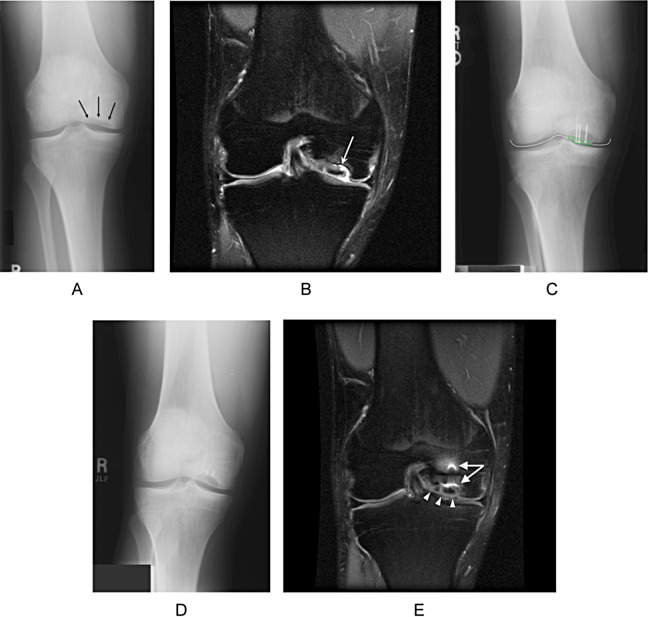
Patient 5. (A) Preoperative anteroposterior (AP) radiographs demonstrating the osteochondritis dissecans lesion of the medial femoral condyle (black arrows). (B) Coronal proton-density-weighted MRI with fat suppression demonstrates the fragment in situ with fluid signal extending to its undersurface (white arrow). The fragment has a similar signal intensity to normal articular cartilage. At the time of surgery, the fragment was found to be completely disconnected from the underlying crater. (C) AP radiograph 1 week after surgery, showing the mini-fragment screws capturing the articular fragment. The radiograph has been graphically enhanced to show the outline of the radiolucent articular fragment (dashed green line) and demonstrate countersinking of the screws beneath the articular surface (solid white line). (D) AP radiograph 9 months postoperatively. At the time of surgery, the cartilage fragment was found to be thicker than normal articular cartilage. Consequently, bone grafting required to reestablish articular congruity did not reestablish the normal subchondral contour, giving the appearance of a persistent defect. The knee had normal joint spaces and no degenerative changes in the medial compartment. Residual screw tips broken off during removal of hardware were buried within the femoral condyle and caused no sequelae. (E) Coronal proton-density-weighted MRI with fat suppression 9 months after surgery, demonstrating the healed cartilage fragment that has completely filled the defect, has a smooth surface, and is congruent with the remaining portion of the medial femoral condyle (white arrowheads). Metal artifact is visible from the residual hardware (white arrows).

