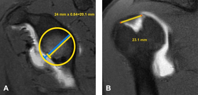
Figure 2.
(A) The glenoid track is calculated as 84% of the actual glenoid width measured on the sagittal oblique magnetic resonance (MR) image. A best-fit circle is placed on the glenoid to calculate the expected width prior to bone loss. Therefore, both percentage of bone loss and glenoid track can be determined. In this case, the actual glenoid width is 24 mm, with 4 mm of bone loss (17% bone loss). The glenoid track is 84% of 24 mm, or 20.1 mm. (B) The distance from the rotator cuff footprint to the medial margin of the Hill-Sachs lesion is measured on the coronal MR. In this case, it is 23.1 mm. Since the Hill-Sachs width to the footprint (23.1 mm) is greater than the glenoid track measurement (20.1 mm), it is considered outside the glenoid track and at high risk for engaging.

