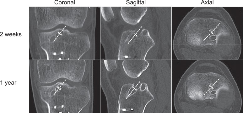
Figure 3.
Computed tomographic images of the knee with anatomic double-bundle anterior cruciate ligament reconstruction at 2-week (top row) and 1-year (bottom row) follow-up. Sclerotic lines of the tibial anteromedial tunnel wall are enhanced in coronal (left), sagittal (center), and axial (right) views.

