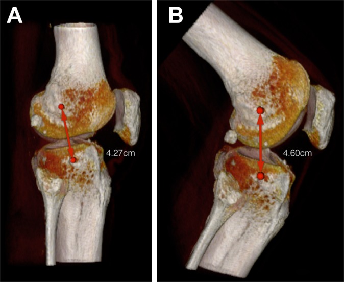
Figure 3.

Three-dimensional computed tomography scans of one of the knees used in the study under (A) full extension and (B) 90° of flexion. Metal markers show an increase in length between the points of origin and insertion of the anterolateral ligament.
