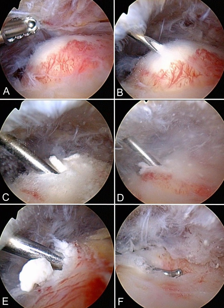
Figure 1.
Operative technique. (A) Partial subacromial bursectomy is performed in the suspected region of calcific deposit (CD) localization (left shoulder). The CD appears as a bump as a result of swelling of the affected supraspinatus tendon. (B) A needle is used to locate the center of the deposit. (C) A blunt hook probe is inserted into the center of the deposit without incising the tendon. (D) “Squeezing” and (E) “stirring” with the hook probe effectuates blunt elimination of carbonate apatite. (F) After CD removal, an indentation is noted at the site of the former bump.

