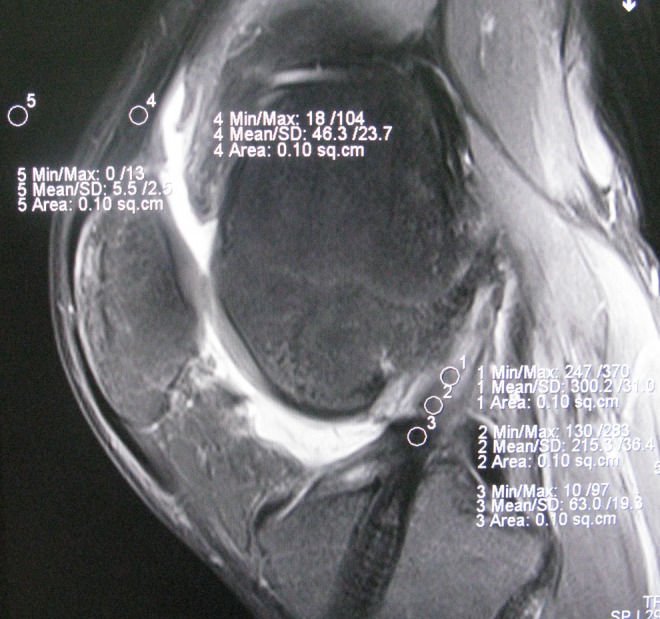
Figure 1.

Sagittal magnetic resonance image of the knee shows the positions of the 5 regions of interest (area of the circle, 0.10 cm2), which included the (1) distal third, (2) middle third, (3) proximal third, (4) quadriceps tendon, and (5) background site (approximately 2 cm anterior to the quadriceps tendon).
