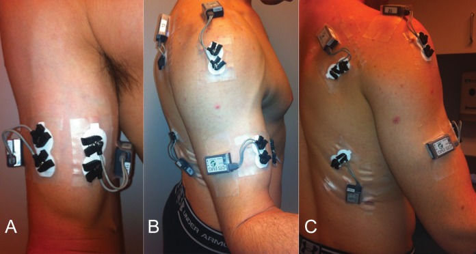
Figure 1.
This series of clinical photographs shows electrode placement. (A) Anterior view demonstrating electrode placement on the long and short heads of the biceps. (B) Lateral view demonstrating electrode location on the middle head of the deltoid. (C) Posterior view showing electrode placement on the infraspinatus. A latissimus dorsi electrode is also shown, although this electrode was not used for this particular study.

