
Figure 5.
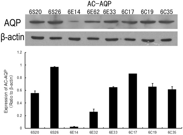
Mitochondrial expression of AQP-8, assessed 6 hours post-surgery using the Western blot technique. β-actin was used as a loading control. The upper panel shows representative immunoblots for AQP8 and β-actin proteins. The lower panel shows data for the sham (6S20 and 6S26), CLP (6C17, 6C19 and 6C35) and CLP+EP (6E14, 6E32 and 6E33) groups, normalized to the β-actin loading control.