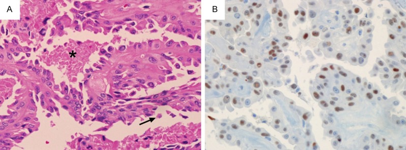
Figure 3.

A. Histology of hobnail variant of papillary thyroid carcinoma. Thin arborizing papillae are lined by hobnail cells. Single detaching cells (arrow) and tumor necrosis (asterisk) are also noted (H&E × 200). B. Immunohistochemistry reveals positivity for p53 (× 200).
