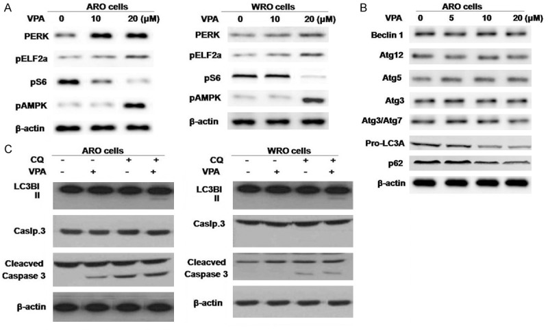
Figure 3.

The effects of VPA on ER stress, metabolic stress, and autophagy. A. ARO and WRO cells were treated with VPA for 48 hours and subjected to Western blot analysis with antibody against molecular markers of ER stress and metabolic stress. B. Western blot demonstrating expression of autophagy markers in ARO cells treated with VPA for 48 hours. VPA induced expression of the lower migrating band of LC3B (LC3B-II) and increased the ratio of LC3B-II (16 kDa) vs. LC3BI (18 kDa). p62 expression was inhibited. C. MTC cells were treated with VPA 10 μM for 48 hours, with or without CQ.
