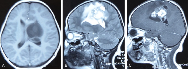
Figure 1.

MR images disclosed a lesion in the left frontal-parietal lobe. A. MR images showed hypointense signal on T1-weighted MR images (axial). B. T2 revealed a large heterogeneous mass lesion in the right frontal lobe (sagittal). C. The central part of the lesion was observed on Gadolinium-enhanced T1 weighed MRI (sagittal).
