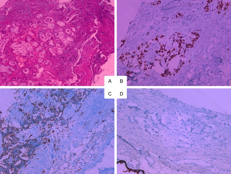
Figure 2.

Pathology slides of the biopsy specimen with different stains. A. Hematoxylin and eosin stained section of the mass demonstrating atypical epithelial cells arranged as irregular glandular or tubular structures infiltrated in the background of proliferative fibrous tissue (×100); B. Immunohistochemical detection of thyroid transcription factor 1 (×100). C. Immunohistochemical detection of napsin A (×100); D. Immunohistochemical detection of CK5/6 (×100).
