
Figure 2.
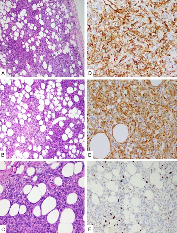
Histological features of fat-forming solitary fibrous tumor (SFT) in the kidney. A. The tumor appeared as a well-circumscribed mass with a fibrous capsule, HE×100; B. The classic appearance of SFT with hemangiopericytoma-like vasculature admixed with clusters or lobules of mature adipose tissue, HE×100; C. Mitotic figure in spindle tumor cells, HE×200; D. Tumor cells were positive for CD34, IHC×200; E. Tumor cells were positive for bcl-2, IHC×200; F. Ki-67 expression was approximately 2% of tumors, IHC×200).