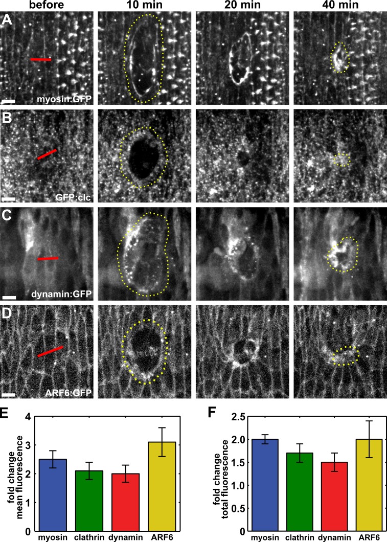
Figure 1.
Components of the endocytic machinery accumulate at the wound margin. (A–D) Epidermal cells expressing myosin:GFP (A), GFP:clc (B), dynamin:GFP (C), or ARF6:GFP (D) in stage 14–15 embryos. Time after wounding is shown. Red lines indicate wound sites. Yellow dotted lines outline the wounds. Anterior left, dorsal up. Bars, 5 µm. (E and F) Maximum fold change in mean (E) and total (F) fluorescence of myosin:GFP (n = 6), GFP:clc (n = 5), dynamin:GFP (n = 7), and ARF6:GFP (n = 6) at the wound margin with respect to the values before wounding. Error bars denote SEM.

