Figure 3.
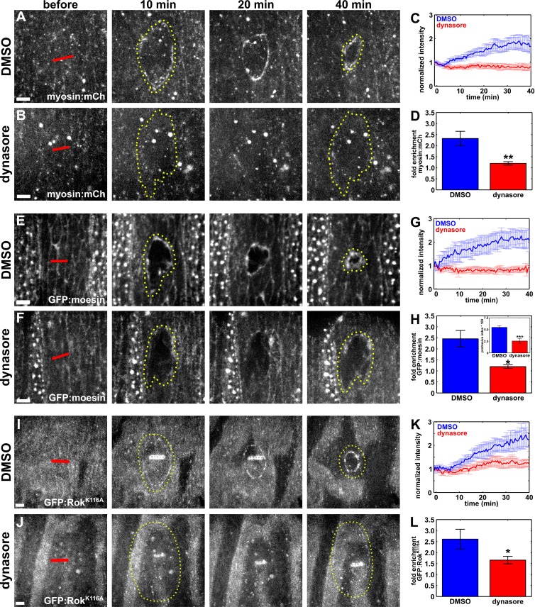
Blocking endocytosis causes defective cytoskeletal remodeling during wound repair. (A, B, E, F, I, and J) Epidermal cells expressing myosin:mCherry (A and B), GFP:moesin (E and F), or GFP:RokK116A (I and J) in embryos injected with DMSO (A, E, and I) or dynasore (B, F, and J) immediately before wounding. Time after wounding is shown. Red lines indicate wound sites. Yellow dotted lines outline the wounds. Anterior left, dorsal up. Bars, 5 µm. (C, G, and K) Mean myosin:mCherry (C), GFP:moesin (G), or GFP:RokK116A (K) fluorescence at the wound margin over time, in control, DMSO-injected embryos (blue; n = 12 in C, n = 5 in G, and n = 6 in K) or dynasore-injected embryos (red; n = 11 in C, n = 5 in G, and n = 8 in K). (D, H, and L) Maximum fold enrichment at the wound margin of myosin:mCherry (D), GFP:moesin (H), or GFP:RokK116A (L). (H, inset) Quantification of protrusive activity at the wound margin. Error bars, SEM; *, P < 0.05; **, P < 0.01; ***, P < 0.001.

