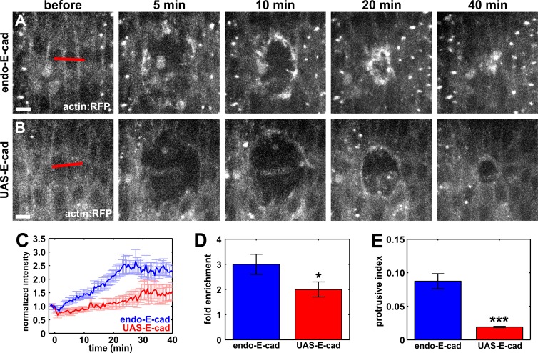
Figure 8.
E-cadherin overexpression causes defects in actin dynamics at the wound margin. (A and B) Epidermal cells in embryos expressing actin:RFP (A and B) and E-cadherin at wild-type levels (A) or overexpressing E-cadherin:GFP (B). Time after wounding is shown. Red lines indicate wound sites. Anterior left, dorsal up. Bars, 5 µm. (C) Actin:RFP fluorescence at the wound margin in embryos expressing wild-type levels of E-cadherin (blue; n = 7) or overexpressing E-cadherin:GFP (red; n = 8). (D) Maximum fold enrichment of actin:RFP at the wound margin. (E) Quantification of protrusive activity at the wound margin. Error bars, SEM; *, P < 0.05; ***, P < 0.001.

