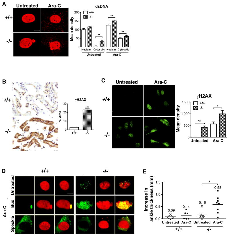
Figure 2. Damaged DNA in the cytosol of Dnase2a-deficient cells.
(A) Fluorescent staining of MLFs from wild-type or Dnase2a−/− mice with anti-dsDNA antibodies; scale bar, 10 μm. Right panel, quantitation of dsDNA in nuclear and cytosolic compartments based on 5 total fields of 20X from 3 independent experiments.
(B) Immunoperoxidase staining of anti-γ-H2AX in kidneys of wild-type and Dnase2a−/− mice, scale bar, 10 μm; right panel, quantitation of signals as % total area based on 5 fields of 20X from 3 matched pairs of wild-type and Dnase2a−/− mice.
(C) Immunofluorescent staining of anti-γ-H2AX in MLFs, scale bar, 50 μm; right panel, quantitation of signals as mean density based on 5 fields of 20X from 3 independent experiments.
(D) Double-staining with anti-γ-H2AX (green) and anti-dsDNA (red) antibodies in MLFs; yellow indicates co-localization; scale bar, 10 μm; images representative of 3 independent experiments.
(E) Increase in ankle thickness from week 0 to week 8 (for both ankles) in matched 10–12 weeks old wild-type and Dnase2a−/− mice (n=3–4), untreated or injected with 3 doses of 15 μg/g Ara-C i.p., mean increase is shown numerically.
See also Figure S2.

