
Figure 1. Kir4.1 and GLT-1 protein expression are reduced in spinal cord of AxD mice.
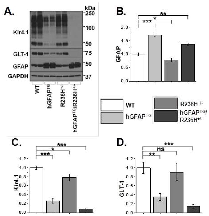
A. Western blot analysis of spinal cord from PND 24–28 WT, hGFAPTG, R236H+/−, and hGFAPTG/R236H+/− mice for Kir4.1, GLT-1 and GFAP and for GAPDH as a loading control. B–D. Quantification of western blot in (A) for GFAP (B), Kir4.1 (C), and GLT-1 (D), normalized to GAPDH levels and WT values. Data are ± SEM, n=4 in all groups, unpaired t-tests, *=p<0.05, **=p<0.01, ***=p<0.001.