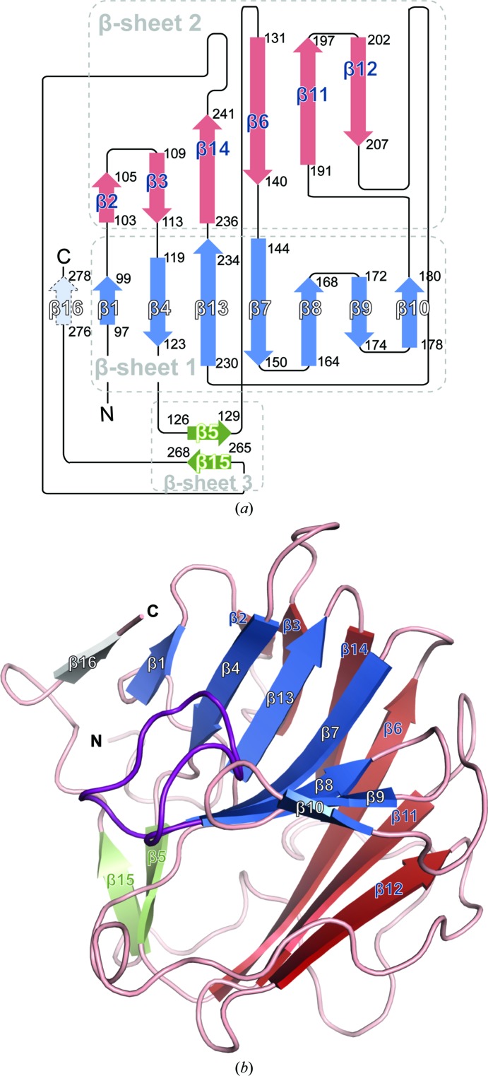
Figure 2.
Structure and topology of hDSPRY. (a) Topology map with β-sheet 1 coloured blue, β-sheet 2 red and β-sheet 3 green. β-Strands are illustrated as arrows. The artificial β-addition module, β-strand 16, of chain B is shown in grey. (b) The β-sandwich fold of hDSPRY; colouring is similar to that in (a). Loop D is highlighted in purple.

