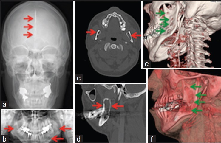
Figure 1.

Images of a patient with Gorlin-Goltz syndrome: (a) Calcification of the falx cerebri ( ). (b) Lesions (
 ) of the mandible. (c) Mandibular cystic lucencies (
 ). (d) Considerable loss of bone (
 ) in the mandible. (e and f) Mandibular odontogenic keratocysts (
 )
)
