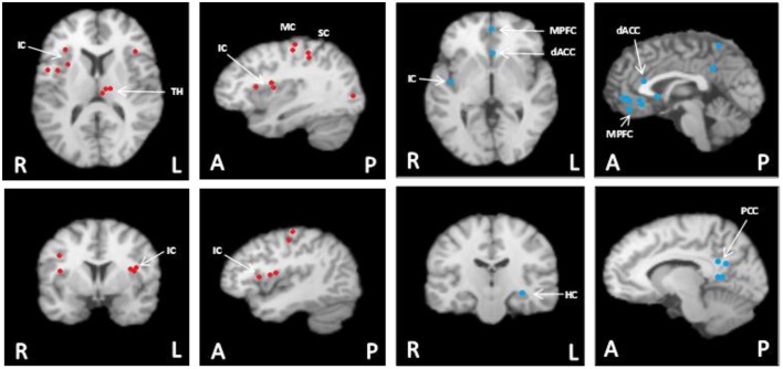
Figure 1.
Meta-analysis summary of data from our laboratory (n = 124) (Wong et al., 2007a,b; Al-Otaibi et al., 2010; Goswami et al., 2011; Norton et al., 2013) of common cortical regions associated with heart rate control during non-fatiguing handgrip exercise task [this figure originally published in Shoemaker et al. (2015) (used with permission)]. These participants each performed 3–7 bouts of moderate intensity (35–40% maximal strength) handgrip tasks each lasting 30 s. Left panels with red dots: Cortical areas of increased activation relative to baseline in response to short duration, moderate intensity isometric handgrip exercise. Right panels with blue dots: Cortical areas of decreased activation relative to baseline in response to short duration, moderate intensity isometric handgrip exercise. FDR pN = 0.01; Min Volume (mm3) = 200. Analysis performed using GingerALE (Version 2.3.2; BrainMap) and Mango (Version 3.1.2; Research Imaging Institute, University of Texas Health Science Center) (Eickhoff et al., 2011; Turkeltaub et al., 2012). MPFC, medial prefrontal cortex; IC, insula cortex; dACC, dorsal anterior cingulate cortex; PCC, posterior cingulate cortex; MC, motor cortex; SC, sensory cortex; TH, thalamus; HC, hippocampus. Images are in radiological presentation with right side of the brain (R) on left, and left side of the brain (L) on the right. A, anterior; P, posterior.

