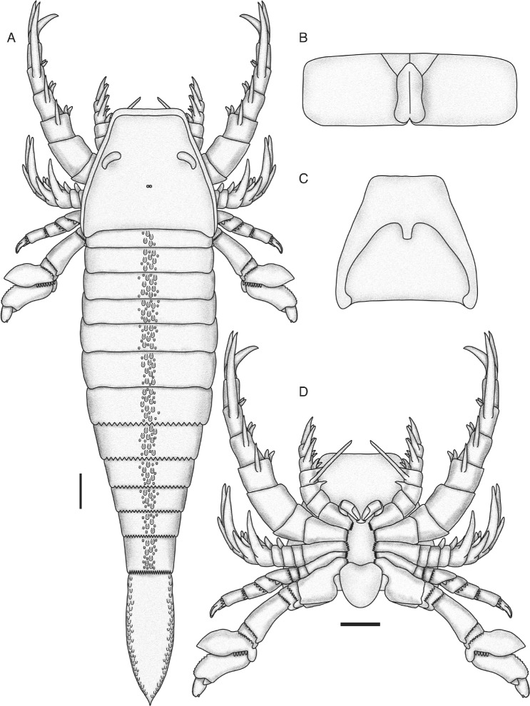
Fig. 20.

Pentecopterus decorahensis, reconstruction of adult. a Dorsal view. b Genital operculum. c Ventral view of carapace and prosomal ventral plate. d Ventral view of prosoma. The appendages are shown rotated in lateral view; in life, appendages IV–VI would be oriented so that the anterior edge (in IV) or the posterior edge (in V and VI) of the limb as in the reconstruction would face ventrally. The form of the median and lateral eyes and metastoma are hypothetical and based on ancestral state reconstructions using the phylogenetic matrix and topology. Scale bars = 10 cm (maximum size)
