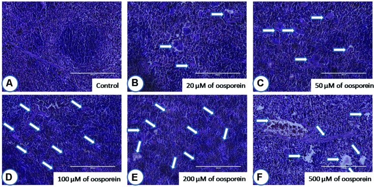
FIGURE 7.
Histopathological observations of spleen upon treatment with different concentrations of oosporein. Histopathology of spleen stained with hematoxylin-eosin-methylene blue. (A) Control spleen showed no histopathological damage, whereas oosporein exposed spleen showed clear evidence of histopathological damage with splenic granulomas, macrophage infiltration and splenomegaly as shown as arrow marks. Damage was graded as (B) mild, (C) mild, (D) moderate, (E) moderate, and (F) severe extreme.

