
Figure 3.
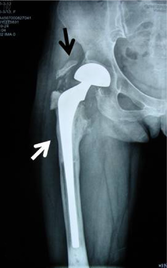
The radiograph shows an 8-year postoperative AP of a cemented allograft prosthesis composite for a chondrosarcoma (IA).
Notes: The white arrow indicates the resorption site; the black arrow indicates the nonunion of trochanter.
Abbreviation: AP, anterior-posterior X-ray.