Fig. 3.

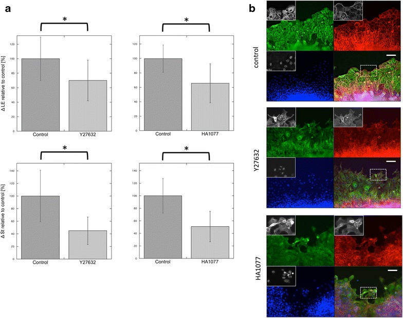
The effect of ROCK inhibitors on the collective cell migration of stratified TE-10 cells. a The distances of migration of cells found along the wound edges for each experimental condition (ΔLE) are shown in the top panels (data shown represent the mean ± SEM). Two inhibitors of ROCKs, Y27632 and HA1077, caused a significant reduction in migration (*p < 0.001). The lower graphs show the distances of migration for cells in the stratified region (ΔSt). Both inhibitors had a statistically significant influence on ΔSt (*p < 0.001). The data show the ratio of the migration distance relative to the control. All experiments were performed at least three times. b Images of the wounded edges of each cell sheet after various experimental treatments. Top control study. The cells in the front row were regularly arranged and well spread. Middle Cells treated with 5 µM Y27632. Many of the cells in the front row showed elongated shapes, with some protrusion of spikes. The arrangement of these cells appeared to be irregular, and the cell sheets were fragmented. Bottom Images of the cells found along the wounded edge after treatment with HA1077. Some of the cells in the leading row were small and round, while others were elongated and similar to the cells treated with Y27632. Empty space was observed between cells, a phenomenon that was not seen in control experiments. Green α-tubulin, red β-actin, blue nuclei; scale bar 100 µm. Each inset shows the magnified image of the area surrounded by the interrupted white line on the merged images
