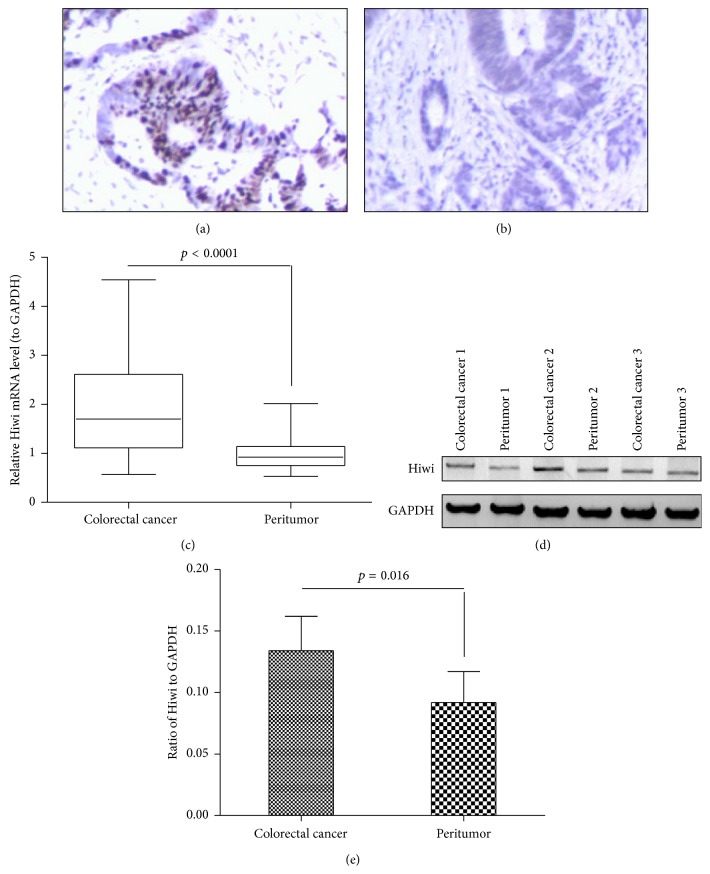
Figure 1.
Overexpression of Hiwi protein in CRC specimens. (a) and (b): immunohistochemical staining for Hiwi expression in CRC tissues (n = 38) and peritumor specimens (n = 38); (c): relative mRNA level of Hiwi in the CRC specimens or the colorectal noncancer specimens by quantitative real-time RT-PCR; (d): Hiwi overexpression at protein level in the CRC or noncolon specimens, revealed by the western blot analysis; (e): percentage of Hiwi to GAPDH in protein level. The p value was indicated accordingly.

