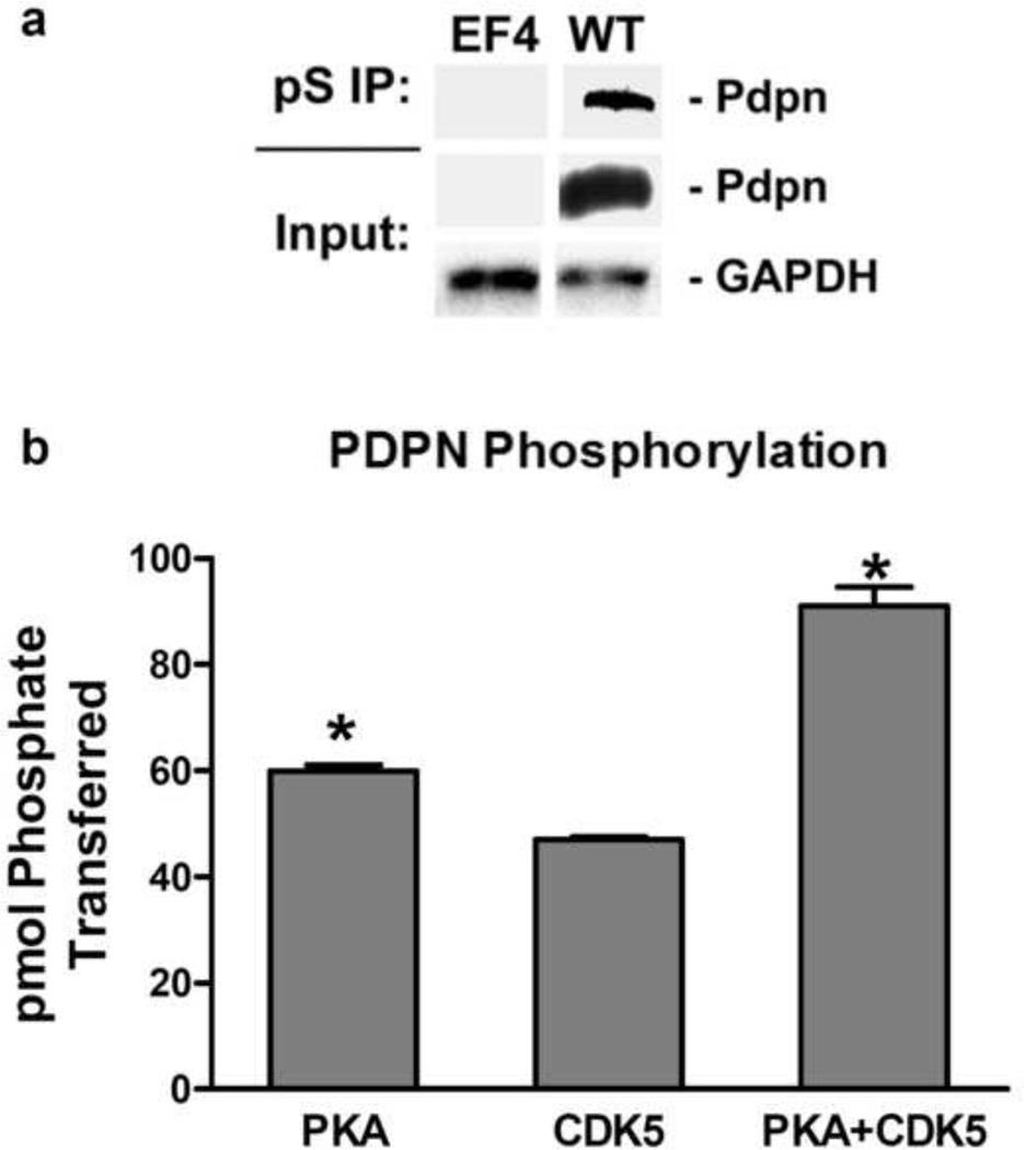
Figure 1. PKA and CDK5 phosphorylate serine residues in the PDPN intracellular domain.
(a) Total protein (input) and protein immunoprecipitated with phosphoserine antiserum (pS IP) from homozygous null PdpnKo cells transfected with empty parental vector (EF4) or wild type PDPN (WT) was analyzed for PDPN and GAPDH by Western blotting as indicated. (b) Peptide containing the entire intracellular region (VVMKKISGRFSP) of PDPN was incubated with PKA, CDK5, or both PKA and CDK5 along with [γ-32]ATP for 10 minutes. Data are shown as picomoles of phosphate incorporated into the PDPN peptide (mean+SD, n=2). Asterisks indicate p<0.05 compared to CDK5 treated cells by t-Test.

