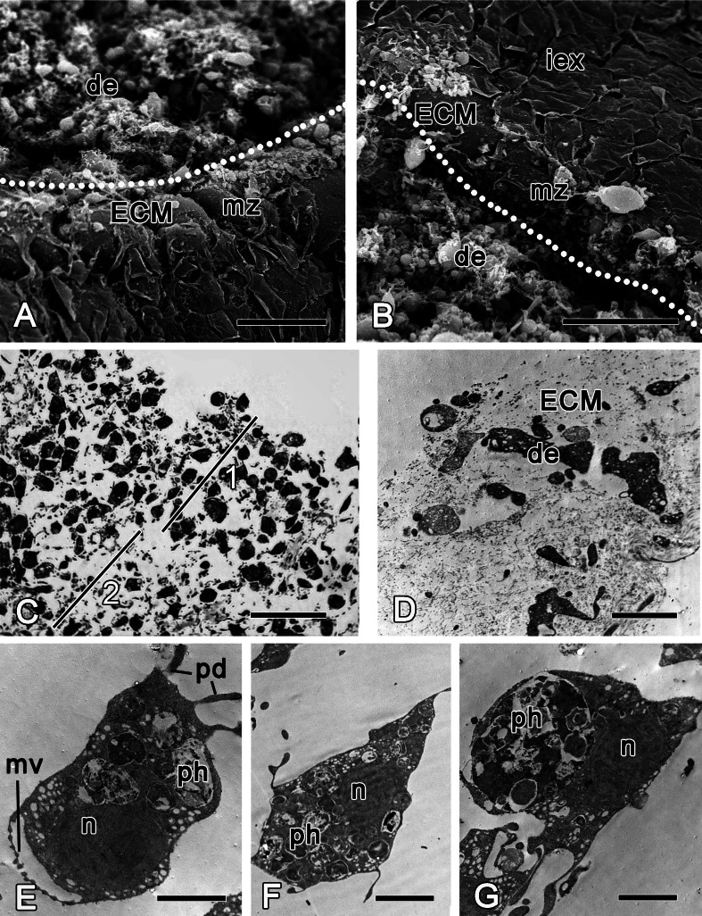
Figure 3. Halisarca dujardini 6 h after injury.
(A) SEM of a wound surface (show with dotted line) with debris (de) and marginal zone, surrounding a wound with dense ECM of the cortex and the exopinacocytes. (B) SEM of intact exopinacoderm, marginal zone and the wound (show with dotted line), covered with debris. (C) Semi thin section of wounded ectosome (1) and adjacent area of choanosome (2). (D) TEM of external part of the wound with cell debris and dense ECM. (E) TEM of dedifferentiating choanocyte in the wound area. (F) TEM of amoebocyte of mesohyl, filled with phagosomes. (G) Endopinacocyte from the wound area filled with big phagosomes. Scale bars: A, B—30 µm; C—25 µm; D–G—2 µm. de, debris; ECM, extra cellular matrix; iex, intact exopinacoderm; mv, microvilli; mz, marginal zone; n, nucleus, pd, pseudopodia; ph, phagosome.

